SECM101: Anatomy of an SECM probe
Latest updated: October 29, 2026Intro
The probe is integral to all scanning probe electrochemistry techniques, Scanning Electrochemical Microscopy (SECM) is no different. In this article the SECM probe is introduced, with its key characteristics defined.
The probe
In an SECM experiment the probe is an UltraMicroElectrode (UME), Fig. 1, held in close proximity to the sample surface. In dc-SECM a dc potential bias is, typically, applied to the UME probe to measure the current of a redox mediator, and thereby map the activity of a sample. In ac-SECM on the other hand, an ac potential bias is applied to measure the impedance of the sample. Not just any UME can be used to perform an SECM experiment, instead consideration must be given to the diameter, shape, and material of the active region of the electrode, as well as the RG ratio, as will be defined later.

Figure 1: A representative SECM probe is shown.
Diameter
The diameter of the SECM probe is particularly significant because it defines the resolution of the final SECM measurement, as compared in Fig. 2. The probe should be selected to be of a similar scale to, or smaller than, the feature of interest. When features are well spaced, the probe can detect features down to one-tenth or even smaller than its diameter [1]. However, to achieve such resolution the features must be well spaced, the probe must be in very close proximity to the sample, and the activity must be strong enough, sometimes driven by biasing the sample, meaning such resolution is typically only expected for the most ideal samples.
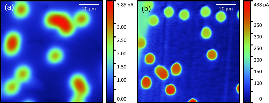
Figure 2 : Generator-collector measurement performed on a polycarbonate membrane with 12 µm pores over a gold sample performed with a (a) 10 µm and (b) 1 µm probe.
Shape
The shape of the active region of the SECM probe is important because it affects mass transport to the probe, in turn influencing the effect of the tip to substrate distance on the signal [2,3], and the image contrast. The majority of SECM measurements are performed using a flat inlaid disc electrode. Aside from being an easily reproducible shape [4], for which much of the SECM theory relates to, an inlaid disc is used because the insulating layer blocks diffusion of the redox mediator to the active region of the probe, when in close proximity to the sample surface, resulting in images with good contrast [5].
Material
While the majority of SECM experiments are performed using platinum [6], other materials, for example gold and carbon, are also used. Materials other than platinum may be used when the redox mediator of interest has a redox potential outside of the electrochemical window available to platinum, or when the redox mediator has an unfavorable, or limited, interaction with platinum.
RG ratio
The RG ratio is the ratio of the insulating sheath of the SECM probe, typically glass, to the active area of the electrode, defined in Eq. 1, and illustrated in Fig. 3. Ideally an RG ratio of 10 or smaller is used, though for the smallest probe sizes larger RG ratios may be acceptable to make the probe more robust. Smaller RG ratios are favored because they make the measurement more sensitive because of increased diffusion of the mediator to the active region of the probe. This increased diffusion allows the probe to approach closer to the sample surface. If the RG ratio is too small, however, the diffusion of the mediator to the active region will not be blocked sufficiently over insulating regions, in particular at the boundary with conducting regions, removing the high contrast expected. If the insulating sheath is completely removed to result in a highly protruding cone, then no contrast is achieved in feedback mode SECM [5].
$$\frac{R}{r} = \mathrm{RG} \leqslant 10 \tag{1}$$
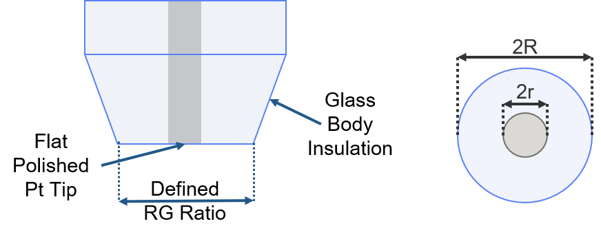
Figure 3 : The RG ratio is illustrated.
Is the probe OK?
Before performing an SECM experiment the probe can be quickly assessed by performing Cyclic Voltammetry (CV) on it far away from the sample surface, i.e. in bulk electrolyte, an example is given in Fig. 4. Although the CV will vary depending on the probe diameter and redox mediator, including its concentration, there are some general characteristics which can be assessed. The CV should have the general sigmoid, or S, shape expected of a UME. If the CV deviates from this shape, for example it is featureless, or has peaks like those seen for a macroelectrode, the probe should be polished. If the CV is a flat, sometimes noisy line, the setup should be checked, this can imply a missing electrode connection, or a poor current range selection, for example. If the CV shows the correct shape, it should also be confirmed that the current measured by the probe is sensible for its diameter with the concentration of redox mediator. This can require some previous knowledge of the probe and mediator behavior. If there is ever a doubt when using a new redox mediator, BioLogic supplies a test sheet with all SECM probes showing their CV behavior with the exact test conditions and solutions listed.
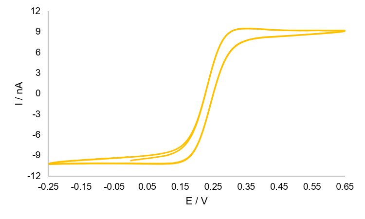
Figure 4 : An example CV, with the characteristic sigmoid shape, measured at a 25 µm Pt SECM probe in 2.5 × 10⁻³ mol/L K3[Fe(CN)6] and 2.5 × 10⁻³ mol/L K4[Fe(CN)6] in 0.1 mol/L KCl. The reference electrode was Ag/AgCl, and the counter electrode was Pt. Measurements were performed from 0.65 to -0.25 V, at a scan rate of 50 mV/s.
Biologic’s SECM probes
BioLogic offers a range of characterized Pt SECM probes covering 1 to 25 µm. All of BioLogic’s SECM probes are tested by cyclic voltammetry, using a standard solution and controlled experimental protocol, to ensure the magnitude and shape of the response are as expected for the diameter of the probe. The result of this test is supplied with the probe. Please note that it is always best practice to polish the SECM probe, even brand-new probes, before use.
References:
- J. Bard, M. V. Mirkin, P. R. Unwin, D. O. Wipf, J. Phys. Chem. 96 (1992) 1861–1868
- J. Bard, F.-R. F. Fan, J. Kwak, O. Lev, Anal. Chem. 61 (1989) 132-138
- Lee, S. Amemiya, A. J. Bard, Anal. Chem. 73 (2001) 2261-2267
- V. Mirkin, F.-R. F. Fan, A. J. Bard, J. Electroanal. Chem. 328 (1992) 47-62
- Skylar, T. H. Treutler, N. Vlachopoulos, G. Wittstock, Surf. Sci. 597 (2005) 181-195
- Polcari, P. Dauphin-Ducharme, J. Mauzeroll, Chem. Rev. 116 (2016) 13234−13278